Breast cancer is a malignant tumor of the breast, most often seen in women.
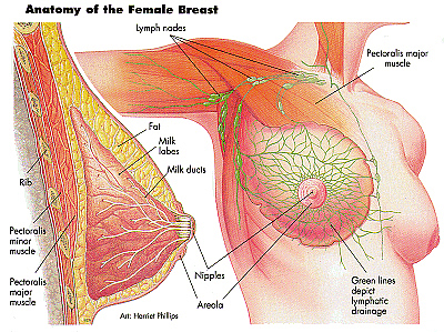
Breast cancer most often begins in the duct lining or in the lobule lining. As you can see from the above picture, the breast is infiltrated with lymph nodes. These lymph nodes connect to the surrounding axillary lymph nodes (arm pits), internal mammary nodes (deep chest), and the supra- or infraclavicular nodes (collarbone). Because of this lymphatic drainage system, cancer can easily spread throughout the body (metastasize). Therefore it is imperative to know if the cancer has spread in order to guide treatment.
Early Detection is Key!
It is advisable that every woman under the age of 40 have a professional breast exam done at least once every three years. For women over 40, a professional breast exam should be performed once a year. Also starting at 40, a woman should start having a mammogram every other year. Beyond the age of 50, a mammogram should be a yearly event. FOR ALL WOMEN: a monthly self breast exam should be done.

***Note: mammograms, even though they use radiation, the doses are so low they do not significantly increase the risk of breast cancer. The amount of radiation emitted is similar to the amount of radiation encountered if one would fly from New York to California on a commercial jet.
Risks
Women who are over the age of 50 are most at risk. Those with a genetic predisposition have their risk factor increase by 80%. Those with a family history are also at a much higher risk. Caucasian women develop breast cancer more, but African American women have a higher mortality rate. Those of Asian, Hispanic or Native American descent have a lower risk of developing breast cancer. Other risk factors include: previous breast radiation, abnormal breast biopsy, atypical hyperplasia, early menarche, not having children, using birth control for more than ten years, using hormone replacement therapy (HRT), high alcohol consumption, obesity with a high fat content around the waist, a high fat diet and sedentary lifestyle.
Symptoms
The most common sign is a lump or mass in the breast. Lumps and masses that are more often cancerous are those that are painless, hard and have irregular edges, though lumps that are tender, soft and round can also be malignant. Check with a doctor if you have any new lumps! There can also be swelling, skin irritation or dimpling, nipple pain or retraction, a red area, scaly skin, or abnormal lactation.
Diagnosis
Common diagnostic tools include mammogram, biopsy, breast ultrasound, ductogram, full field digital mammography, computer aided detection and diagnosis, scintimammography, MRI, nipple discharge exam, ductal lavage, nipple aspiration, chest X-ray, CT scan, and PET scan. These tests are used for diagnosis but some can also be used to measure invasiveness, progression, metastasis, prognosis and grade of the cancer.
There are different grading systems for cancer. Histological grade is used for invasive types of cancer. The lower the grade, the slower the cancer is growing. It measures the arrangement of cells in relation to each other. Grade One shows well differentiated cells that look normal, grow slowly and the tubular structure is small. Grade Two shows moderately differentiated cells that show features of cells between Grades One and Three. Grade Three shows poorly differentiated cells that lack normal features that are growing and spreading aggressively.
The nuclear grade of a tumor depicts how abnormal cells are and the presence or absence of necrosis. There are also tests done that look for estrogen or progesterone receptors to determine if the cancer is hormonally dependent.
The TNM grading system is also used:
- Primary Tumor
- TX – primary tumor cannot be assessed
- T0 – no evidence of primary tumor
- Tis – Carcinoma in situ – interductal, lobular, Paget’s
- T1 – tumor <=2 cm
- T2 – tumor >2cm/<5cm
- T3 – tumor >5cm
- T4 – any size with extension to chest wall or skin
- Regional Lymph Nodes (N)
- NX – lymph nodes cannot be assessed
- N0 – no regional lymp nodes
- N1 – metastasis to movable ipsilateral axillary lymph nodes
- N2 – metastasis to ipsilateral axillary lymph nodes fixed to one another/structures
- N3 – metastasis to ipsilateral internal mammary lymph nodes
- Distant Metastasis (M)
- MX – distant metastasis cannot be assessed
- M0 – no distant metastasis
- M1 – distant metastasis, including supraclavicular nodes
| Stage 0 | Stage I | Stage IIA | Stage IIB | Stage IIIA | Stage IIIB | Stage IV | |
| TNM | TisN0M0 | T1N0M0 | T0-1N1M0
T2 |
T2N1M0
T3N0M0 |
T3N1M0
T0-3N2M0 |
T4, any N/T, N3M0 | Any T/N, M1 |
| 5yr Survival Rate | 92% | 87% | 78% | 68% | 51% | 42% | 13% |
Types of Breast Cancer
Adenocarcinomas incorporate almost all breast cancers. Adenocarcinoma refers to cancer of a glandular tissue and is a term used most often when describing cancers of the lung, stomach and breast.
| Type | Origin | Description |
| Non Invasive
(In Situ) |
Ductal | (20%) Cancer cells limited to the duct and have not spread. Found through mammograms and are easily cured. If necrosis is present, the cancer is more aggressive. |
| Lobular | Not a true cancer. Limited to milk producing glands. Not invasive but increases the risk of developing invasive breast cancer. | |
| Invasive
(Infiltrating) |
Ductal | Most common type (80%). Starts in milk passage or duct and invades the fatty tissue of the breast, where it can spread through lymph system, blood system, or metastasize. |
| Lobular | (10%) Start in milk producing glands or lobules. It is harder to detect through mammogram and tends to metastasize. |
Other types of breast cancer include: inflammatory breast cancer, medullary carcinoma, mucinous carcinoma, Paget’s disease of the nipple, Phyllodes tumor, and tubular carcinoma.
Breast Cancer according to Traditional Chinese Medicine
Breast Cancer in TCM is know as Fu Yan or ‘Chest Rock.’ It is generally first diagnosed using western medicine methods such as mammograms, X-rays, ultrasounds, etc. Symptoms often include chest swelling that is hard and fixed and generally in a single breast, nipple secretion, nipple shape change (flattening, depression, erosion), or skin change.
| Syndrome | Breast Symptoms | Other Symptoms | Formula |
| Disharmony of Chong and Ren | Hard, fixed swelling in the chest, unsmooth surface | Five center heat, afternoon fever, night sweating, dry mouth, sore low back and knees, irregular menstruation, red cracked tongue, less tongue coating, thready (rapid) and weak pulse | Modified Zhi Bai Di Huang Wan |
| Liver Qi Stagnation | Distending pain in the chest and hypochondriac region | Depression or anxiety, bitter taste in mouth, dry throat, dizziness, vertigo, thin and white/yellow tongue coating, slightly dark tongue, taut and smooth pulse | Modified Xiao Yao San |
| Toxic Heat Stagnation | Fast growing swelling in the chest with pain and redness, maybe broken skin with foul liquid | Constipation, fever, dark red tongue, taut fast pulse | Modified Tao Hong Si Wu tang |
| Qi & Blood Deficiency | Swelling(s) in the chest that are fixed and hard to move, stick to chest wall, bumps on chest | Dizziness, vertigo, pallid complexion, fatigue, less tongue coating, pale tongue, toothmarked tongue, weak pulse | Modified Bu Zhong Yi Qi Tang |
Commonly used herbs for breast cancer include Shan Ci Gu, Feng Fang, Zi Cao, Ai Ye, Tian Men Dong.
Caroline Prodoehl, D.Ac.
References;
Zhu, Anni Yawen (2011). Integrated Treatments: Internal Medicine Course. TorontoSchoolof Traditional Chinese Medicine. Toronto,Ontario.